🔬 VLab4Mic explores this in silico, connecting molecular models with super-resolution experiment design.
#Microscopy #SuperResolution #VLab4Mic @aiopticalbiolab.bsky.social
 @dctomaz.bsky.social
 @dctomaz.bsky.social
🔬 VLab4Mic explores this in silico, connecting molecular models with super-resolution experiment design.
#Microscopy #SuperResolution #VLab4Mic @aiopticalbiolab.bsky.social
 @fabmusacchio.bsky.social
🗣️ The 6th Day of Mouse Intravital #Microscopy 🔬 will take place at our institute @dzne.science and you can still register:
⏰ Abstracts: Sep 24, 2026
📅 Nov 23-24, 2026
🌍 https://gerbi-gmb.de/event/6th-day-of-intravital-microscopy/

 @mycologycorner.bsky.social
Lyophyllum eustygium
#fungifriends #microscopy #mycology #mushroomsky #shroomscrolling


 @kentmochizuki.bsky.social
肝臓の組織構造です。
過去の研究でラットから採取したHE染色切片像を倍率ごとに並べています。
肝臓に運ばれた血液は種々の細胞の働きにより老廃物の除去、解毒や代謝などのお手入れを受けます。
その構造・機能美に感心します。
Zoom into the liver from the slice to cells, in a HE-stained rat liver thin section.
Thickness: 3μm (paraffin)
🔬Olympus, BX51 (4x, 10x, 20x, 40x)
@mycologycorner.bsky.socialPaxillus obscurisporus
#fungifriends #microscopy #mycology #mushroomsky #shroomscrolling


 @thepurplelilac.bsky.social
Happy Fluorescent Friday!
Under the microscope:
Green (β3-tubulin): Retinal ganglion cells cytoskeleton
Blue (DAPI): Cell nuclei
A simple yet beautiful look at retinal cellular architecture.
#FluorescentFriday #Microscopy #Neuroscience #RGC #Sciart #ScienceCommunication #Stemcells #Organoids
 @mycologycorner.bsky.social
Parasola kuehneri
#fungifriends #microscopy #fungifriday #mushroomsky #shroomscrolling


 @mariescopy.bsky.social
The #microscopy community has serious concerns about this year’s winning #NikonSmallWorld in Motion video.
@healthcare.nikon.com, could you please comment on the raw data and acquisition process to clarify whether AI was used? Transparency would help maintain confidence in the competition.
 @rt-superes.bsky.social
Live lattice-SIM reveals lysosome repair as a spatial event: DFCP1-positive ER microdomains dynamically assemble around damaged lysosomes and help concentrate lipid-transfer machinery for repair.
 @astrobrian.bsky.social
When a laser beam hits an electron beam
#microscopy #ElectronMicroscopy #cryoEM #cryoET #microSky #scienceSky
 @museumofscience.bsky.social
Most people's morning starts with a cup of coffee. Ours started with a cup of coffee under a microscope. ☕🔬
Our friend Chloé Savard, also known as tardibabe on Instagram, zoomed in and found a whole universe brewing in there.
 @so-lets-kilab70.bsky.social
Yesterday Cherry Huang gave us a friendly reminder at our Seminar Series: your fixative is not an innocent bystander
She did an amazing job “schooling” us how crosslinking chemistry alters biosensor localization in #chromatin #microscopy
Great science, beautiful slides & impressive #scicomm skill
 @mycologycorner.bsky.social
Hortiboletus bubalinus
#fungifriends #microscopy #mushroomsky #shroomscrolling


 @healthcare.nikon.com
🦠 Celebrating #InternationalMicroorganismDay!
Don't miss the 2026 Nikon Small World in Motion 2nd Place winner: "A tiny roundworm and a single-celled Dileptus explore their microscopic world" by Nguyen Nam Nhat.
🎥 Watch the full video: https://bit.ly/3T6TWKO
 @kill-bait.bsky.social
Nikon Small World in Motion 2026 Winners Reveal Hidden Microscopic Life
🤖 IA: It's not clickbait ✅
👥 Users: It's not clickbait ✅
#microscopy #nikonsmallworld #scientificimaging
👇👇👇
 @uniofnomi.bsky.social
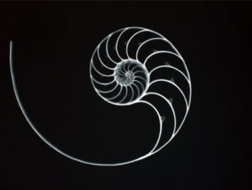
Here's a scanning electron #microscope image of an avian (bird) #schistosome larva penetrating human skin and causing "swimmer's itch."
Check out all the spikes decorating its forked tail! OUCH!!
(Photo credit: Curtis L Blankespoor)

 @microscopy.confocalnl.com
Great to be at @embo.org #EMBOCLEM! 🔬
Today, attendees used our AION confocal microscope for a CLEM experiment, enabling rapid four-color imaging and direct correlation of fluorescence data with EM images.
Great to see AION in action!


 @leah.myatproto.social
@washuengineers.bsky.social 's Song Hu has developed an amazing microscopy tool for #neuroscience research. Put it to work, #ScienceSky 🧪#NeuroSky 🧠 #Microscopy 🔬
source.washu.edu/2026/09/oxyg...
 @visitech.bsky.social
We’re at the Francis Crick Institute this week, supporting the Multi-dimensional Imaging and Analysis microscopy training course.
If you’re attending, come over and say hi, and find out more about fast, live-cell super-resolution imaging with the VT-iSIM.

 @moritz-negwer.mstdn.science.ap.brid.gy
Thursday 24th September, 11:30 - 17:00 at de Exponent, ground floor of the research tower of the RIMLS Nijmegen: https://maps.app.goo.gl/mE3ZjbEz71BD54nu7